Приказ основних података о документу
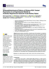
Immunohistochemical Pattern of Histone H2A Variant Expression in an Experimental Model of Ischemia–Reperfusion-Induced Acute Kidney Injury
| dc.creator | Nešović-Ostojić, Jelena | |
| dc.creator | Životić, Maja | |
| dc.creator | Kovačević, Sanjin | |
| dc.creator | Ivanov, Milan | |
| dc.creator | Brkić, Predrag | |
| dc.creator | Mihailović-Stanojević, Nevena | |
| dc.creator | Karanović, Danijela | |
| dc.creator | Vajić, Una Jovana | |
| dc.creator | Miloradović, Zoran | |
| dc.creator | Jovović, Đurđica | |
| dc.creator | Radojević-Škodrić, Sanja | |
| dc.date.accessioned | 2023-06-16T10:47:02Z | |
| dc.date.available | 2023-06-16T10:47:02Z | |
| dc.date.issued | 2023 | |
| dc.identifier.issn | 1661-6596 | |
| dc.identifier.uri | http://rimi.imi.bg.ac.rs/handle/123456789/1295 | |
| dc.description.abstract | Ischemia–reperfusion injury (IRI) is a frequent cause of AKI, resulting in vasoconstriction, cellular dysfunction, inflammation and the induction of oxidative stress. DNA damage, including physical DNA strand breaks, is also a potential consequence of renal IRI. The histone H2A variants, primary H2AX and H2AZ participate in DNA damage response pathways to promote genome stability. The aim of this study was to evaluate the immunohistochemical pattern of histone H2A variants’ (H2AX, γH2AX(S139), H2AXY142ph and H2AZ) expression in an experimental model of ischemia–reperfusion-induced acute kidney injury in spontaneously hypertensive rats. Comparing the immunohistochemical nuclear expression of γH2AX(S139) and H2AXY142ph in AKI, we observed that there is an inverse ratio of these two histone H2AX variants. If we follow different regions from the subcapsular structures to the medulla, there is an increasing extent gradient in the nuclear expression of H2AXY142ph, accompanied by a decreasing nuclear expression of γH2AX. In addition, we observed that different structures dominated when γH2AX and H2AXY142ph expression levels were compared. γH2AX was expressed only in the proximal tubule, with the exception of when they were dilated. In the medulla, H2AXY142ph is predominantly expressed in the loop of Henle and the collecting ducts. Our results show moderate sporadic nuclear H2AZ expression mainly in the cells of the distal tubules and the collecting ducts that were surrounded by dilated tubules with PAS (periodic acid–Schiff stain)-positive casts. These findings may indicate the degree of DNA damage, followed by postischemic AKI, with potential clinical and prognostic implications regarding this condition. | |
| dc.relation | info:eu-repo/grantAgreement/MESTD/inst-2020/200015/RS// | |
| dc.relation | info:eu-repo/grantAgreement/MESTD/inst-2020/200110/RS// | |
| dc.rights | openAccess | |
| dc.rights.uri | https://creativecommons.org/licenses/by/4.0/ | |
| dc.source | International Journal of Molecular Sciences | |
| dc.subject | AKI | |
| dc.subject | H2AZ | |
| dc.subject | histone | |
| dc.subject | immunohistochemistry | |
| dc.subject | ischemic–reperfusion injury | |
| dc.subject | γH2AX | |
| dc.title | Immunohistochemical Pattern of Histone H2A Variant Expression in an Experimental Model of Ischemia–Reperfusion-Induced Acute Kidney Injury | |
| dc.type | article | |
| dc.rights.license | BY | |
| dc.citation.issue | 9 | |
| dc.citation.spage | 8085 | |
| dc.citation.volume | 24 | |
| dc.identifier.doi | 10.3390/ijms24098085 | |
| dc.identifier.fulltext | http://rimi.imi.bg.ac.rs/bitstream/id/2942/Immunohistochemical_patternt_of_histone_H2A_pub_2023.pdf | |
| dc.type.version | publishedVersion |